An AI-Driven Workflow to Improve Macula Detection and Retinal Ganglion Cell Quantification in Preclinical NHP Histopathology

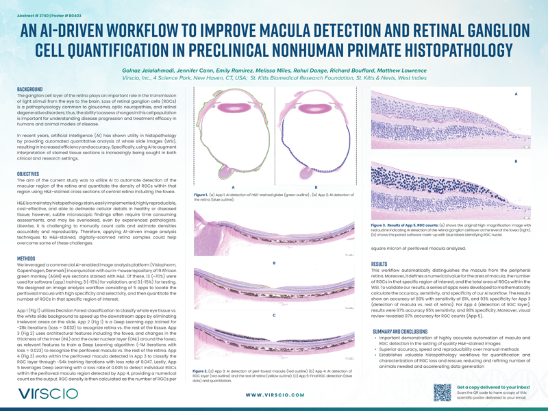
This poster presents an innovative approach using artificial intelligence (AI) to enhance the detection and quantification of the macula and retinal ganglion cells (RGCs) in hematoxylin and eosin (H&E) stained whole slide images (WSI) from nonhuman primate tissues. Our research highlights the integration of advanced AI techniques into preclinical histopathological assessments, a pivotal step forward in understanding and diagnosing retinal diseases such as glaucoma.
At Virscio, we leverage the findings from this research to enhance our preclinical research offerings, providing more accurate and reproducible data for therapeutic development. The application of AI in our histopathological processes allows us to deliver detailed and precise analyses, facilitating the advancement of drug development strategies and supporting our partners in bringing innovative therapeutics to market.
In this scientific poster, you will learn more about:
- Learn about the application of AI in histopathological assessments of nonhuman primate tissues
- Understand the challenges and solutions in the detection and quantification of the macula and retinal ganglion cells
- Discover the benefits of automated histopathological evaluations, including improved accuracy and efficiency
- Explore the implications for drug development and therapeutic innovations in the field of retinal diseases
Ready to Deepen Your Understanding?
Download Our Scientific Poster Now!
